Introducing MammoWave®: an innovative medical device for breast cancer detection
HOW IT WORKS
Introducing MammoWave®: an innovative medical device for breast cancer detection
DISCLAIMER
This section must be accessible only to healthcare professionals, it will not be visible to the general public (according to local regulations on MDR advertise)
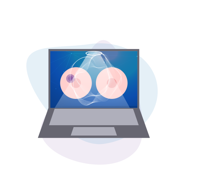
It employs a novel technique which generates images by processing very low power (<1 mW) electromagnetic signals in the microwave band (1-9 GHz).
The MammoWave Device
MammoWave is the medical device involved in the project clinical investigation. Il employs a novel technique which generates images by processing very low power (<1 mW) electromagnetic signals in the microwave band (1-9 GHz) .
The development of the technique
The development of microwave breast cancer detection technique has been driven by the dielectric properties contrast between cancers and healthy breast tissues. This contrast is shown to be up to a factor of five in conductivity and permittivity. Meanwhile, newer studies suggest the existence of such contrast only between healthy fatty and malignant breast tissues, and a lower contrast between healthy fibro glandular and malignant tissues.
MammoWave® is composed of two antennas which illuminate the breast tissue using electromagnetic fields in the microwave band and measures the correspondent scattered electromagnetic fields.
The software
The technology is supported by a proprietary software augmented by Artificial Intelligence (AI) built around a dedicated imaging algorithm. Images are dielectric parameters’ homogeneity maps, shown in 2D, azimuthal plane (i.e. coronal plane).
The software is augmented by Artificial Intelligence (AI) and calculates some parameters (such as the ratio between the maximum and the average intensity). The software automatically provides a dedicated label indicating the presence or absence of suspicious findings in each breast.
Join our Community
Find out the latest news about MammoScreen world and stay up to date with the project.
This section must be accessible only to healthcare professionals, it will not be visible to the general public (according to local regulations on MDR advertise)