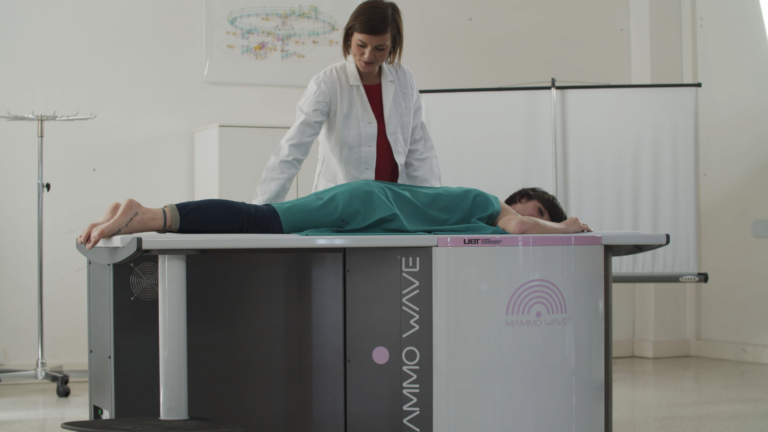
MammoWave works differently from traditional methods that use X-rays.
The device generates a qualitative representation of the breast tissues by processing very low power (<1 mW) electromagnetic signals in the microwave band (1-9 GHz).
The ability to use this range of the electromagnetic spectrum for breast cancer detection is based on the difference in the dielectric properties of healthy and affected tissues. In simple terms, healthy and affected tissues respond differently to electromagnetic fields in the microwave band which the MammoWave device is able to contrast and display.
The exam takes a few minutes per breast and is performed with the patient lying in a comfortable facing-down position. MammoWave allows for a totally discreet examination, improving the compliance of patients when going for regular breast screening.

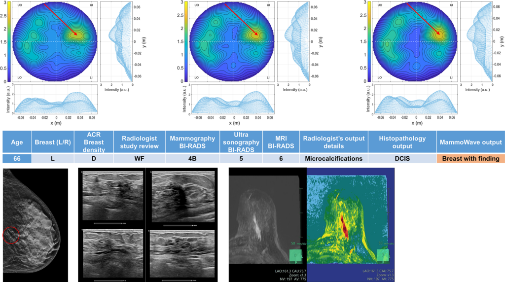
The results obtained using MammoWave are displayed as dielectric properties homogeneity maps shown in two dimensions, crossing a coronal plane (i.e., separating the front and back of the body in an imaginary plane that cuts through both shoulders).
The signals are analysed with a proprietary software that uses a novel, fast and accurate algorithm to highlight a lack of homogeneity in the breast. The software calculates certain parameters (such as the ratio between the maximum and the average intensity) and is augmented with Artificial Intelligence (AI) to help identify suspected lesions.

MammoWave has several important advantages when compared to the classic X-ray mammography. One is that the microwave range used in the scan is completely safe. Another is that it does not require compression of the breast, which makes the process more comfortable for the person being examined.
This article is part of #1 issue of MAMMOSCREEN newsletter: read the full issue here.


